Abdominal Pain in Pediatric Emergency

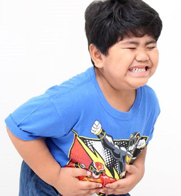
Causes of abdominal pain in childhood Abdominal pain is one of the more common reasons for parents to bring their child to the Emergency Department. While many diagnoses traverse all age groups, some are more age specific (Table 1).

Abdominal Pain in Pediatric Emergency


Assessment of abdominal pain

The assessment of the child with acute abdominal pain depends on a good history and careful examination. The nature of the pain itself must be carefully elicited, including its characteristics, relieving and precipitating factors. Truly severe colicky pain suggests an obstruction of the gastrointestinal, genitourinary or hepatobiliary tract. Pain can be referred from elsewhere such as the testes and the lungs. Associated features must also be elicited such as gynecological symptoms in adolescent girls. A full and careful but sensitive examination must follow. If acute activity precipitates pain, it suggests peritonitis. Acute serious problems need rapid combined surgical and medical assessment and management. The algorithm (Table 1) may be used as a guide to the systematic consideration of various categories of causes of acute abdominal pain. Typical features of some important causes of acute abdominal pain in children are described in the following table (Table 2).

Notes:
• Acute appendicitis must be considered in any child with severe abdominal pain, aggravated by movement such as walking, or a bumpy car ride. The child is often flushed, with a tachycardia and a mildly elevated temperature. In the very young child, in whom the risk of perforation is higher, the presenting symptoms are less specific. The diagnosis is clinical—no laboratory or radiological tests are required, although there is usually an elevated white cell count.
• The peak age for intussusception is 6–12 months. The child may present in shock. Plain anterior chest X-ray may show signs of bowel obstruction, with decreased gas in the right colon. The diagnosis is confirmed by air insufflation or barium enema, with reduction usually possible by the same means (unless there are signs of peritonitis, which increase the risk of perforation).
• Mid-gut volvulus is commonest in the newborn period, but can occur in later childhood. Predisposing factors include malrotation and abnormal mesentery.
• Vomiting is rarely due to constipation.
• Some children suffer recurrent non-specific abdominal pain, with no organic cause identifiable. Constipation is often an important contributing factor. Psychogenic factors (e.g. family, school issues) need to be considered. These children should be referred for general pediatric assessment.
• Some less common diagnoses need to be considered in patients with certain underlying chronic illnesses. Hirschsprung disease can be complicated by enterocolitis, with sudden painful abdominal distension and bloody diarrhea. These patients can become rapidly unwell with dehydration, electrolyte disturbances, and systemic toxicity, and are at risk of colonic perforation. Primary bacterial peritonitis can occur in children with nephrotic syndrome, splenectomy and those with VP shunts.

Causes of abdominal pain by age
Table 1.  Causes of abdominal pain by age

Assessment of abdominal pain in pediatric
Table 2. Assessment of abdominal pain in pediatric

Management

An algorithm for abdominal pain management is given in Table 1.
Intravenous access should be established. The electrolytes should be measured in a child who appears dehydrated and blood and stool cultures obtained if the child is potentially septic. The patient should be given and fasted until a surgical opinion has been sought. In addition, a nasogastric tube will be needed if there is bowel obstruction.
Meningitis in Pediatric Emergency

The presentation of a child with meningitis varies with age. Infants with meningitis frequently present with non-specific signs and symptoms such as fever, irritability, lethargy, poor feeding and vomiting. The fontanelle may or may not be full. Older children may complain of headache or photophobia. Neck stiffness may be present (although this is not a reliable sign in young children). A purpuric rash is suggestive of meningococcal septicemia. It is important to examine for spinal and cranial abnormalities such as dermal sinuses, which may have predisposed the child to meningitis.

Note: This guideline is not for use in children with spinal abnormalities or ventriculoperitoneal shunts where the neurosurgical team should be consulted.

Meningitis in Pediatric Emergency


Assessment

Assess airway, breathing and circulation. Monitor pulse, blood pressure, respiratory rate, oxygen saturation and consciousness. Insert intravenous line and take blood for glucose, FBC and blood cultures and U&Es, meningococcal PCR, and CRP (C-reactive protein). Do nose and throat swabs, as occasionally the organism is isolated from these. If the clinical diagnosis is meningitis and there are contraindications for a lumbar puncture give intravenous antibiotics without delay.

Lumbar puncture (LP)
An LP is used to confirm the diagnosis of bacterial meningitis and to identify the organism and its antibiotic sensitivities. In some cases it should be postponed because of the risk of coning. There are contraindications to performing an LP, including:
• Coma—Glasgow Coma Scale (GCS) <13 or rapid deterioration in conscious state or absent or non-purposeful responses to painful stimuli (squeeze earlobe hard for up to 1 minute. Children should localise response and seek a parent)
• Focal neurological signs
• Papilledema
• Cardiovascular compromise
• Evidence of coagulopathy.

If any of these signs are present the patient is in danger of coning, and management in Intensive Care is usually required. Notify a PICU immediately (for retrieval or transfer), perform blood cultures, commence antibiotics.

Remember cerebrospinal fluid (CSF) findings in early bacterial meningitis may mimic a viral pattern or even be normal (see Table 6.1). In a traumatic tap, allow 1 white blood cell for every 500 red blood cells, and 0.01 gl−1 of protein for every 1000 red cells. Request culture and analysis for bacterial antigens (this is non-urgent as it does not change immediate management and there is no need for it to be performed out of hours).


Management

a. Admission to a PICU
Admission to PICU should be discussed with the consultant in the following circumstances:

Meningitis: Factors requiring consultant opinion
•Age less than 2 years
•Coma
•Cardiovascular compromise
•Intractable seizures
•Hyponatremia

b. Antibiotics
In a child aged over 2 months the usual organisms causing bacterial meningitis are Streptococcus pneumoniae, Neisseria meningitidis and Hemophilus influenzae type b (HiB—uncommon after the age of 6 and the incidence is reduced following HiB vaccination).

Empirical treatment consists of cefotaxime (50 m g/kg weight per dose up to a maximum of 3 g, 6 hourly) given prior to determination of the organism responsible and its sensitivities. Chloramphenicol may be used in children with a type 1 hypersensitivity to cephalosporins. You may need to consider the addition of vancomycin if pneumococcus is suspected in an area of high incidence of penicillin-resistant pneumococcus.

Continue empirical treatment until cultures are known to be negative or an organism and its sensitivity pattern are known. A positive culture result with sensitivities should lead to narrower spectrum treatment.

In a child aged under 2 months, the organisms to consider in this age group include group B streptococcus, Escherichia coli and other Gram negative organisms, Listeria monocytogenes, Streptococcus pneumoniae, Neisseria meningitidis and Hemophilus influenzae type b.

Initial therapy is with i.v. benzylpenicillin (50mg/kg weight per dose, 6 hourly), and i.v. cefotaxime (50 mg/kg weight per dose, 6 hourly), and i.v. gentamicin (dose according to age). Ongoing therapy is modified according to culture and sensitivity results. Consult local guidelines.


Treatment in meningjtis with positive cultures with sensitivities
  • Neisseria meningitidis—give i.v. benzylpenicillin 50 mg/kg weight per dose up to a maximum of 3 g, 4 hourly for 7 days (in penicillinsensitive cases)
  • Streptococcus pneumoniae—give i.v. benzylpenicillin 50 mg kg−1 per dose up to a maximum of 3 g, 4 hourly for minimum of 10 days (in penicillinsensitive cases)
  • Hemophilus influenzae—give i.v. cefotaxime 50 mg/kg weight  per dose up to a maximum of 3 g, 6 hourly for 7–10 days, or i.v. amoxycillin 50 mg/kg weight  per
  • dose 4 hourly for 7–10 days (depending on sensitivities)
  • Other—if an organism is not isolated, but significant CSF pleocytosis is present, a minimum of 7 days treatment with i.v. cefotaxime is recommended.


c. Fluid management
Careful fluid management is important in the treatment of meningitis as many children have increased antidiuretic hormone (ADH) secretion. The degree to which fluid should be restricted varies considerably from patient to patient depending primarily on their clinical state.

Shock should be corrected with 20 ml/kg weight of normal saline. A patient who is not in shock and whose serum sodium is within the normal range should be given 50% of maintenance fluid requirements as initial management. If the serum sodium is less than 135 mmol/l give 25–50% of maintenance requirements. The serum sodium should be repeated every 6–12 hours for the first 48 hours and the total fluid intake altered accordingly. If the serum sodium is less than 135 mmol/l, reduce the fluid intake.

d. General measures
• Neurological observations including blood pressure should be performed every 15 minutes for the first 2 hours and then at intervals determined by the child’s conscious state.
• Weight and head circumference should be monitored on a daily basis.
• Control seizures.
• Early consultation with intensive care unit is necessary for any child who is experiencing a deterioration in conscious state, hemodynamic instability or seizures.
• Electrolytes should be checked every 6–12 hours until the serum sodium is normal.
• Ensure adequate analgesia (e.g. paracetamol) for children in the recovery phase who may have significant headache.
• The role of steroids is still controversial and we have opted generally not to use them, although they may be indicated in Hemophilus meningitis.


Isolation

Children with meningococcal disease require isolation until they have had 24 hours treatment. Other children with meningitis can be nursed on the open ward.

Fever persisting for more than 7 days

This may be due to nosocomial infection, subdural effusion or other foci of suppuration. Uncommon causes include inadequately treated meningitis, a parameningeal focus or drugs.


Contact chemoprophylaxis

Practice differs in different countries. Below is one regime used in Australia. It is important that prophylaxis be given early to both the index case and contacts as follows:
• Index Case and all household contacts if household includes other children under 4 years of age who are not fully immunized.
 Index Case and all household contacts in households with any infants under 12 months of age, regardless of immunization status.
• Index Case and all household contacts in households with a child 1–5 years of age who is inadequately immunized.
• Index Case and all room contacts including staff in a child-care group if Index Case attends over 18 hours per week and any contacts under 2 years of age who are inadequately immunized. (NB. Inadequately immunized children should also be immunized.)
• Index Case (if treated only with penicillin) and all intimate, household or day-care contacts who have been exposed to the Index Case within 10 days of onset.
• Any person who gave mouth-to-mouth resuscitation to the Index Case.

Australian contact chemoprophylaxis regimen
  • Hemophilis influenzae type b—give rifampicin 20 mg/kg weight orally as a single daily dose to a maximum 600 mg) for 4 days. For infants <1 month give rifampicin 10 mg/kg weight  orally daily for 4 days. In pregnancy or contraindication to rifampicin give ceftriaxone 125 mg/kg weight  i.m. (<12 years) or 250 mg/kg weight  i.m. (>12 years) as a single dose.
  • Neisseria meningitidis—give either: rifampicin 10 mg/kg weight  orally 12 hourly up to a maximum 600 mg for 2 days. For infants <1 month give rifampicin 5 mg/kg weight  orally 12 hourly for 2 days. In pregnancy or contraindication to rifampicin give ceftriaxone 125 mg/kg weight  i.m. (<12 years) or 250mg/kg weight  i.m. (>12 years) as a single dose. or ciprofloxacin 500 mg orally as a single dose.
  • Streptococcus pneumoniae—there are no increased risks to contacts, so no antibiotic required.
  • Rifampicin may cause orange-red discoloration of tears, urine and contact lenses, skin rashes and itching, and gastrointestinal disturbance. It negates the effect of the oral contraceptive pill and should not be used in pregnancy or severe liver disease.


Headaches in Pediatric emergency

Headache is a common symptom in children, affecting 80–90% by the age of 15. The common causes are systemic illness with fever, local ENT problems, migraine and tension headache. Meningitis, raised intracranial pressure (ICP), e.g. from tumors, and subarachnoid hemorrhage (SAH) are much rarer causes but these need to be considered. Any headaches that wake a child from sleeping or that are associated with focal signs such as a hemiplegia require investigation as an inpatient

Headaches in Pediatric emergency


Assessment

History
It is useful to classify headache as acute or recurrent. The following list gives the causes and key features to help make a diagnosis, based on careful history and examination:

Acute
• Systemic: fever and general illness (e.g. ’flu, pneumonia, septicemia)
• Local sinusitis, dental caries, otitis media
• Trauma: head injury
• Meningitis: reduced conscious level, toxicity, photophobia, neck stiffness
• SAH: sudden onset, severe occipital pain; possible reduced conscious level, neck stiffness

Recurrent
• Migraine: aura, nausea, vomiting, pallor, family history
• Tension: throbbing pain (involving neck muscles) at end of day
• Behavioral: family/social/school problems (may be difficult to identify)
• Raised ICP: morning headaches ± vomiting, worse with coughing/sneezing/bending
• Progressively worsening raised ICP: personality or behavioral changes, focal neurological symptoms
• Benign intracranial hypertension, systemic hypertension, uremia, recurrent hypoglycemia, recurrent seizures, lead or CO poisoning.

It may be useful to make out a possible family headache patterns diagram to help identify the nature of the headache.

Examination
As part of the examination it is important to document the following:
• ABC: blood pressure, heart rate
• General: toxic, unwell, temperature, rash
• Neurology: conscious level, fundi, visual fields, cerebellar signs, neurocutaneous stigmata, neck stiffness, cranial bruits
• Local causes: cervical lymphadenopathy, teeth, sinus, ears
• Growth and puberty: head growth, height, weight, growth velocity, pubertal status.

Investigations
In the acute situation, the two most important questions to answer are:
• Does the child need an urgent computerised tomography (CT) scan of the head? and
• Should a lumbar puncture (LP) be performed? In making the decision, you should consider the following factors:
• If the child has altered state of consciousness, focal neurological signs, raised blood pressure or papilledema, consider management of raised ICP and CT scan of head. Discuss this with senior medical staff.
• Consider LP (in the absence of the contraindications) if you are concerned about meningitis or SAH. You may need to do a CT scan first (discuss with consultant).
• If there are no symptoms and signs suggesting raised ICP/SAH/ meningitis and the story is suggestive of migraine, then treat symptomatically (see below).
• If other causes are suspected, do the appropriate investigations (e.g. septic screen, urea, carboxyhemoglobin or lead level [wrist X-rays], blood sugar profile).


Management

If there is a specific diagnosis such as meningitis, SAH, tumor, systemic infection or local infection, then treat as appropriate. Most recurrent headaches can be managed by a pediatrician and do not need to be referred to a neurologist.


Abort attack
Avoid opiates. Initially try simple oral analgesics such as paracetamol (20 mg/kg weight stat then 15 mg/kg weight per dose every 4 hours, to a maximum of 4g per day) or codeine (1 mg/kg weight per dose every 4 hours) or NSAID (ibuprofen 2.5–10 mg/kg weight per dose 6–8 hourly). For adolescents give 1 g of aspirin, 1 g of paracetamol, 10 mg of metoclopramide. Some, but not all, pediatricians use intravenous anti-emetics in severe vomiting. For example, if vomiting is a prominent feature in children over 10 years give slow i.v. prochlorperazine
(0.1–0.2 mg/kg weight).

Prophylaxis

Refer to a local doctor or general pediatric outpatient clinic for long-term management. Consider beta blockers, pizotifen or calcium channel blockers. Non-pharmacological interventions (e.g. avoidance of triggers, relaxation) often play an important role in prevention. Consider getting the child or parents to make a headache diary.

Headache patterns
It may be a good idea with the use of the chart below to explore the pattern of the child’s headaches .
• Acute recurrent includes migraine (common, classical, complicated).
• Chronic non-progressive includes tension (stress related); muscle contraction; anxiety; depression; somatisation headaches.
• Chronic progressive includes headaches from tumor; benign intracranial hypertension; brain abscess; hydrocephalus.
• Acute on chronic non-progressive includes tension headache with coexistent migraine.

Febrile Convulsions

A simple febrile convulsion is a brief (<15 min) generalized convulsion in a febrile (>38°C) child aged between 6 months and 6 years, with no previous afebrile seizures, no progressive neurological condition and no central nervous system infection. Febrile convulsions are common, and occur in 3% of healthy children between the ages of 6 months and 6 years. They are usually associated with a simple viral infection. The onset of the convulsion may be sudden with little evidence of preceding illness. The convulsion may be terrifying for the parents to observe—they frequently believe that their child is dying and may attempt CPR or other resuscitative measures. Febrile convulsions are benign, with minimal morbidity and essentially no mortality. Most febrile convulsions are brief and do not require any specific treatment. The initial management of the convulsion is described in the afebrile seizure section.


After the convulsion

A septic work-up including lumbar puncture is mandatory in children under 6 months of age; by definition a febrile convulsion should not be diagnosed in a child under 6 months. Lumbar puncture should be very strongly considered in those aged 6–12 months. Possible clinical scenarios and management include those shown in Box 6.2. Most children with simple febrile convulsions who have recovered sufficiently do not need to be admitted to hospital If a patient is discharged home following a febrile convulsion, it is important to give the family advice regarding fever control and what to do in the event of a future convulsion. Verbal advice should be reinforced with written advice. Follow up during the next 24 hours is advisable to assess the progress of the child’s illness and to allow parents the opportunity for further discussion.

Possible clinical scenarios and management following febrile convulsions


Repeated convulsions during the same illness occur in about 10–15% of children. The child should be reassessed in hospital. There are usually no serious implications; however, a period of observation might be required to clarify the progress of the illness.


Fever control

Clothing should be minimal—a nappy alone or light outer layer depending on ambient temperature. Tepid sponging, baths and fans are ineffective in lowering core temperature, and are not recommended. Paracetamol has not been shown to reduce the risk of further febrile convulsions. It may be used for pain or discomfort or as an antipyretic associated with febrile illnesses such as otitis media. The parents should understand the reasons for its use and be discouraged from trying to get their child’s fever down.


Long-term issues
Recurrence rate depends on the age of the child—the younger the child at the time of the initial convulsion, the greater the risk of a further febrile convulsion. Epilepsy. Risk of future afebrile convulsions is increased by a family history of epilepsy, any neurodevelopmental problem or complex, very prolonged or focal febrile convulsions. 

Risk of afebrile convulsions following a febrile convulsion
• No risk factors: 1% risk of future epilepsy, similar to the general population
• 1 risk factor: 2% increased risk
• More than 1 risk factor: 10% increased risk

Anticonvulsant treatment. Children who have recurrent prolonged convulsions (which are rare) may benefit from having a rectal diazepam kit available at home, which their parents can administer if a convulsion does not cease spontaneously within 5 minutes. Long-term anticonvulsants are not indicated except in rare situations with frequent recurrences. It may be appropriate to offer a review appointment with a general pediatrician.

Convulsions in Pediatric Emergency

Afebrile convulsions and status epilepticus

Most convulsions are brief and do not require any specific treatment Convulsions may be generalized and tonic-clonic in nature but can also be focal. Generalized convulsive status epilepticus (CSE) is currently defined as a convulsion lasting 30 minutes or more or when successive convulsions occur so frequently that the patient does not recover consciousness between convulsions. Tonic-clonic status occurs in up to 5% of patients with primary epilepsy and about 5% of children with febrile seizures may present with CSE.


Management of the convulsion

The primary assessment should be as for any seriously ill child. A structured approach to the primary survey is particularly important; if a problem is found during the initial ABCDE assessment then immediate treatment and resuscitation should occur,remembering to treat the treatable as you progress through the primary survey. In a fitting child never forget to check the glucose and treat hypoglycemia appropriately. If a convulsion occurs, position the child in a semiprone position to minimise the chance of aspiration. If necessary and possible, clear the airway with gentle suction and give oxygen via the facemask.

If the convulsion continues for more than 10 minutes then follow the flow chart in pict below.
Give high-flow oxygen via facemask, monitor oxygen saturation and test the glucose level. A short acting benzodiazepine should then be administered. The nature and dose of the drug will depend on whether intravenous access had been obtained and on departmental guidelines. Some antiepileptics such as paraldehyde are used in some countries but not others.

Convulsions management and treatment in Pediatric Emergency


a. If venous access or intraosseous access has been obtained
The following drugs can be given in sequence if fitting continues:
• Lorazepam 0.1 mg/kg weight i.v. or intra-osseous (i.o) can be given and repeated after 10 minutes if necessary. This is currently the drug of choice in the latest Advanced Pediatric Life Support Guidelines in the UK, USA and Australia. Diazepam 0.25 mg/kg weight or midazolam 0.15mg/kg weight are alternatives to lorazepam. They can be repeated after 5 minutes if seizures continue. The child needs to be monitored carefully for respiratory depression.
• Paraldehyde 0.4 ml/kg weight per rectum (p.r.) is given 10 minutes after the benzodiazepines if necessary, but is not available in Australia.
• Phenytoin 18 mg/kg weight i.v. or i.o. as a loading dose in normal saline over 30 minutes can then be given with ECG monitoring. If the child is already on phenytoin, then i.v. phenobarbitone 15–20 mg/kg weight can be used over 10 minutes. In neonates, phenobarbitone is often used as the drug of choice.
• If fitting still continues, seek senior anesthetic and medical advice and assistance. The child may require rapid sequence induction and intubation with thiopentone 4 mg/kg weight i.v. or i.o. Muscle relaxants should not be used long term.

b. If venous access has not been obtained
• Diazepam 0.25–0.5 mg/kg weight rectally or midazolam 0.15 mg/kg weight i.v. can be given.
• After 10 minutes, give paraldehyde 0.4 ml/kg weight p.r. (0.4ml of paraldehyde plus 0.4 ml of olive oil=0.8 ml/kg weight of prepared solution), but this is not a choice in Australia.
• Seek medical assistance as above for vascular access and RSI.

Notes
Non-convulsive status may occur. It manifests as decreased conscious state with or without motor accompaniments. It may occur particularly in the Lennox-Gastaut syndrome, or in other children with developmental delay. Pyridoxine-dependent seizures should be considered in any infant under 18 months with recurrent or refractory afebrile seizures. A clinical trial of pyridoxine 100 mg i.v. is warranted. If it is going to be effective it will work within 10–60 minutes. If it is not effective in 10 minutes begin other standard anticonvulsants as above.


After the convulsion

First seizures, even if unprovoked, rarely require continuous anticonvulsant medication. Discuss with the Registrar or a Consultant. Parents should be warned that all children or adolescents who have had seizures should be supervised when bathing, swimming, or riding a bicycle on the road, and that children should avoid tree-climbing. Parents should be advised of first-aid measures should the seizure recur. In some centres, children with their first afebrile seizure are referred to the general pediatric outpatient clinic for follow up. If the child has not fully recovered, or there are concerns about the underlying etiology, he or she should be admitted to hospital.

Coma in Pediatric Emergency

Coma is a symptom, not a diagnosis. The aim of management is to minimise any ongoing neurological damage. History, examination, investigation and treatment will be simultaneous.

Immediate resuscitation and management

The primary assessment should be as for any seriously ill child. A structured approach to the primary survey is particularly important; if a problem is found during the initial ABCDE assessment, then immediate treatment and resuscitation should start, remembering to treat the treatable as you progress through the primary survey.

Supporting an inadequate airway, ineffective breathing or compromised circulation should reduce the likelihood that the reduced conscious level is secondary to hypoxia and fluid loss and of any further deterioration of conscious level due to these two mechanisms. Thus, you must clear and protect the airway and assist the breathing as necessary. If a traumatic cause for the symptoms is likely, immobilise the cervical spine and arrange urgent neurosurgery-involvement.

Check the pulse and insert an intravenous line and perform a blood sugar test; if blood glucose is less than 2.5 mmol 1 −1 in a person who is not diabetic, send specific blood tests  and administer 10% dextrose i.v., 5 ml/kg weight bolus. Consider naloxone, 0.1 mg/kg weight (max 2mg) if pupils are small. Assess and monitor pulse, respiratory rate, BP, temperature, oximetry and conscious state. Consider signs of raised intracranial pressure and look for subtle signs of continuing convulsions.

History and examination

Consider the onset and duration of symptoms. Is there a past history? Relevant conditions include seizures, diabetes, adrenal insufficiency, infection and cardiac problems. Other signs and symptoms may suggest the cause of the coma (Figure 1).

Cause of Coma in Pediatric
Figure 1: Cause of Coma in Pediatric


Investigations and further management

Consider these investigations in the light of the possible diagnoses, if not already done:
• full blood examination
• urea and electrolytes
• glucose
• liver function
• arterial blood gas
• urine drug ± metabolic screen
• urine antigens
• blood and urine culture
• ammonia
• cortisol
• coagulation screen
• ECG

Investigation and management of coma

Figure 2 shows the investigation and management of coma. It is important that raised intracranial pressure is always excluded before lumbar puncture in all cases.

investigation and management of coma in pediatric
Figure 2: investigation and management of coma

Figure 2 Lumbar puncture, imaging and antibiotics in coma. Abbreviations:CT, computed tomography; EEG electroencephalogram; ICP, intracranial pressure; LP, lumbar puncture; MRI, magnetic resonance imaging.


Ongoing care

Continue to assess and support the airway, breathing and circulation. Care will depend on the diagnosis, level of consciousness and degree of ventilatory and circulatory support required. Consider an early transfer to a pediatric intensive care unit.

Pneumothorax in Pediatric Emergency

Definition

A pneumothorax is defined as the presence of air in the intrapleural space. Spontaneous pneumothorax is unusual. It is often associated with trauma but can be spontaneous, particularly if a person has a marfanoid habitus. It can occur secondary to a bullous or a cyst. Children with cystic fibrosis are more at risk of developing pneumothoraces. Consider pneumothorax in a child presenting with pleuritic chest pain or shortness of breath. There may be a previous history of pneumothorax. On examination, there may be reduced air entry and a deviated trachea (away from the collection) and hyper-resonance, but no added breath sounds.

Management

In small pneumothoraces, a conservative approach is appropriate. Where there is less than 30% reduction of total lung capacity (TLC), initial high flow oxygen will help absorb the intrapleural air and in most cases the pneumothorax will resolve over a 1–2 week time scale. Where there is between 30–50% reduction in TLC there are three steps depending on response:
• Oxygen therapy in cases where there are no major symptoms and observation and repeat X-ray over 12 hours.
• Oxygen therapy and a needle thoracocentesis using a small catheter attached to a 3-way tap. This is inserted in the second intercostal space above the rib and in the midclavicular line. Air is aspirated, the tap is closed off and chest X-ray repeated over the following 6 hours. If the air has not reaccumulated, the catheter can be removed and the patient reviewed 24 hours later with repeat X-ray.
• Insertion of an intercostal drain with a valve or underwater seal. This should be placed in the anterior axillary line in the fourth intercostal space.

Pneaumonia in Pediatric Emergency


Pneaumonia in Pediatric Emergency
Pneaumonia in Pediatric Emergency Thorax Xray
Pneumonia is common, but the cause may vary with age. The commonest bacterial causes are:
• Staphylococcus aureus in children <1 year
• Streptococcus pneumoniae in children <4 years
• Hemophilus influenzae in children <10 years
• Group A streptococcus in children >10 years

Viral infection with RSV (respiratory syncytial virus) and parainfluenza occurs in children, especially in those under 2 years of age, and with influenza virus in older children. Mycoplasma infection is more common in older children. In a previously well child older than 1 month, consider pneumonia in infants and children with the following signs and symptoms.

Chest X-ray (AP) should be performed to confirm or exclude pneumonia. Patients with wheeze and air trapping most commonly have bronchiolitis or asthma. Neonates who are unwell, or have a temperature over 38°C, should have a chest X-ray as part of a septic work-up, especially if the respiratory rate is elevated or there are other signs of respiratory embarrassment.

Management of pneumonia

Pneumonia can be managed with inpatient or outpatient care depending on the severity of the condition.

Croup or Barky Cough (Laryngotracheobronchitis)

This tends to occur in a previously well child aged 3 months to 6 years but can occur in older children. The term croup refers to a clinical syndrome characterised by barking cough, inspiratory stridor and hoarseness of voice. It results from viral infection, most often with parainfluenza virus with inflammation of the upper airway, including larynx, trachea and bronchi; hence the term laryngotracheobronchitis.

Croup or Barky Cough (Laryngotracheobronchitis)


The symptoms are typically worse at night and peak on about the second or third night. Differentiating spasmodic croup from viral croup is diffi-cult and often not useful.Consider other causes of acute stridor, such as epiglottitis (much rarer since Hib vaccine), bacterial tracheitis (rare), or laryngeal foreign body (very rare). Refer to upper airways obstruction section.

History

The loudness of the stridor is not a good guide to the severity of obstruction. Children with pre-existing narrowing of the upper airways (e.g. subglottic stenosis, congenital or secondary to prolonged neonatal ventilation) or children with Down syndrome are prone to more severe croup and admission should be considered even with mild symptoms.


General Management

Avoid distressing procedures, e.g. examining throat, because anxiety exacerbates croup. Nurse the child on the parent’s lap. Blood tests, pulse oxymetry, or O2 mask are rarely indicated. A routine nasopharyngeal aspirate (NPA) is not required for children with a ypical clinical picture of croup.

Primary assessment and initial management of croup

1. Mild
Signs: No stridor at rest, Not distressed, No sternal retraction, No signs of hypoxia.
Managements: Can be managed at home. No specific treatment is usually required, although steroids can be considered if the patient is seen early in the course of the illness. Explanation to parent needed

2. Moderate
Signs: Stridor at rest, Distressed, Sternal retraction at rest, Normal breath sounds, No signs of hypoxia
Managements: Administer steroids, Observe and settle child with minimal interventions, Consider use of nebulized epinephrine (adrenaline) (1% eye drop solution 0.05 mg/kg weight or 1:1000 preparation 0.5 ml/kg weight (max 2ml), If settles well after prednisolone consider discharge (see notes on discharge)

3. Severe
Signs: Marked stridor at rest or may be soft, Very distressed, Marked increase work of breathing, Reduced breath sounds, Evidence of hypoxia (restless, lethargy, pallor, cyanosis)
Managements: Continually assess the child and response to therapy, Involve senior staff early on Administer nebulized epinephrine, Administer steroids, Rarely will require intubation, Admission to PICU


Steroids
There is little evidence supporting the use of steroids in mild croup, although the current studies are underpowered. However there is good evidence for the efficacy of corticosteroids for the treatment of moderate to severe croup. There are a number of ways of administering the steroids: use nebulized budesonide, oral prednisolone or dexamethasone or i.m. dexamethasone. Varying doses have been suggested in the literature and all are effective in the treatment of the acute symptoms. Cost and availability may influence the treatment choice. Suggested doses are prednisolone (1 mg/kg weight), nebulized budesonide 2 mg, dexamethasone (0.15 mg/Kg weight).

Epinephrine (adrenaline)
There has been a change in the use of nebulized epinephrine (adrenaline) in the last few years and nebulized epinephrine (adrenaline) no longer needs to be reserved only for children with severe croup. Current evidence would support the use of nebulized epinephrine (adrenaline) in children with moderate and severe croup. In a number of selected children following observation in the Emergency Department for 3 hours after the administering of nebulized epinephrine (adrenaline) and the start of steroid treatment,it may be safe to discharge the child home. Nebulized epinephrine (adrenaline) may cause circumoral pallor.


Admission or discharge

The decision to admit a child is made after initial treatment and observation. As is usual with other children, the time of the day, parent’s anxiety and access to transport, and ability of early review should be taken into account if admission or discharge is being considered. Some centres will suggest that if the child still has stridor at rest after treatment, he or she should be admitted. It has been suggested that if there is no sign of increased work of breathing and no sternal recession but minimal stridor at rest, then they could be discharged with adequate explanation and follow up.

Acute Upper Airways Obstruction

Examination and assessment

A harsh barking cough with stridor in a child with minimally raised temperature suggests croup (see below). Cough with low pitched expiratory stridor and drooling suggests epiglottitis. Sudden onset of coughing, choking, drooling and aphonia suggests a laryngeal foreign body (this is extremely rare). Swelling of the face and tongue with wheeze and urticarial rash suggests anaphylaxis. The differential diagnosis is seen in

Examination

The mouth and throat should not be examined if signs of partial upper airway obstruction are present, as complete obstruction can ensue during the examination. Partial acute upper airways obstruction is characterised by stridor and increased work of breathing. Signs of deterioration are those of hypoxia (worried, restless), fatigue, decreased conscious state, and increased and then decreased work of breathing. Heart rate may be rapid or indeed slow. A child’s general appearance is more useful.

Treatment

Allow the child to settle quietly on the parent’s lap and observe closely with minimal interference. Treat specific cause (see croup/anaphylaxis guidelines). Call the Pediatric Intensive Care Unit (ICU) if the child’s condition is worsening or there is severe obstruction, or call for senior doctors (ENT/anesthetics) if an ICU is not available in your hospital

Oxygen may be given while you are awaiting transfer transport. It can be falsely reassuring because a child with quite severe obstruction may look pink with oxygen. 

Note that:
• Intravenous access should be deferred—upset can cause increasing obstruction.
• Lateral X-rays do not assist in management. In severe airways obstruction, X-rays cause undue delay in definitive treatment and may be dangerous (positioning may precipitate respiratory arrest).
• Do not examine the throat with a tongue depressor.
• If intubation is considered necessary, this should be done by the most experienced medical personnel present, using a smaller than normal endotracheal tube 
This should ideally be done in the operating room with tracheostomy equipment and a surgeon in attendance.
• Once the airway is protected, then full blood count (FBC)/blood cultures, i.v. cannulation and antibiotics can be started.

Bronchiolitis in Pediatric Emergency

This is an acute viral lower respiratory infection. Varying definitions throughout the world cause confusion when doctors are assessing useful treatments. The etiological agent is usually respiratory syncytial virus but can also be adenovirus or parainfluenza 3.

The infection usually affects children under 1 year old. It occurs in about 10% of all infants and 2–3% are admitted to hospital. Younger infants are usually more seriously affected. The illness usually peaks on day 2–3 with resolution of wheeze and respiratory difficulty over 7–10 days. The cough may persist for weeks and postbronchiolitic respiratory symptoms may cause much concern. Management involves a primary assessment of severity and initiation of resuscitation and treatment. Assessment and management are illustrated in Table of Primary assessment and initial management of bronchiolitis in below.

Primary assessment and initial management of bronchiolitis


History

The child presents with cough and wheezing. A lethargic, exhausted child may feed poorly, be hypoxemic, and is at risk of respiratory failure. Risk factors include age, infants with bronchopulmonary dysplasia or congenital heart disease. The time course is important. Is the child improving, stable, or likely to deteriorate over the next few days? Peak severity is usually at around day 2–3 of the illness. If the child is early on in the illness, consider admission to hospital.


Examination

Clinical signs include cough, tachypnea and hyperinflation. There may be audible wheeze. Auscultation reveals widespread crepitations and wheeze and signs of accessory muscle use. Cyanosis always indicates severe disease. Acyanotic infants may also be hypoxemic. If O2 saturation is less than 90%, the infant should receive supplementary O2 during the examination.


Investigations

A routine NPA (nasopharyngeal aspirate) or chest X-ray is not required for children with a typical clinical picture of bronchiolitis. If a chest Xray is taken it may demonstrate hyperinflation, peribronchial thickening, and often patchy areas of consolidation and collapse.

Consider whether this may be early asthma. Nebulized salbutamol may help in the older infant. Currently there is little evidence supporting the use of bronchodilators or antibiotics in bronchiolitis. Steroids were not thought to be of benefit, although some recent work from Toronto suggests that they may help.

Asthma in Pediatric Emergency

Acute asthma is one of the commonest reasons for presentation to an emergency department and admission to a hospital. Consider acute asthma when a child presents with signs of increased work of breathing, widespread wheezing and shortness of breath. There are other causes to consider such as Mycoplasma pneumonia, aspiration, inhaled foreign body, and cardiac failure. In the setting of a child with a previous history of asthma or when asthma seems the most likely diagnosis, then perform a primary assessment of severity and institute the initial resuscitation and treatment.


Primary assessment and initial resuscitation and management

Table below shows the clinical signs and management of asthma at varying degrees of severity. The arterial oxygen saturation (SaO2) may be reduced in the absence of significant airway obstruction by factors such as atelectasis and mucous plugging of airways. SaO2 is purely a measure of oxygenation, which may be preserved in the presence of deteriorating ventilation (with CO2 retention).

Consider transferring the child to a pediatric intensive care unit if the child:
• is in impending respiratory failure
• requires continuous nebulizers for >1 hour
• requires salbutamol more frequently than every 30 minutes after 2 hours
• is becoming exhausted.

Consider ventilation if:
• PCO2 is >8kPa
• there is persistent hypoxemia with PO2<8kPa in inspired oxygen of 60%
• there is increasing exhaustion despite emergency treatment.
Note that exhaustion, a silent chest, cyanosis, bradycardia and hypotension are preterminal signs.


Tables of Assessment and Management of Asthma in Pediatric


Tables of Assessment and Management of Asthma in Pediatric Mild degree
Mild Degree


Tables of Assessment and Management of Asthma in Pediatric Moderate degree
Moderate Degree


Tables of Assessment and Management of Asthma in Pediatric Critical degree
Critical Degree


Tables of Assessment and Management of Asthma in Pediatric Severe degree
Severe Degree

If poor response to i.v. salbutamol give aminophylline 10 mg/kgweight i.v. (maximum dose 250 mg) over 60 min.
Following loading dose, give continuous infusion (1–9 years: 1.1 mg/kgweight per hour, 10+ years: 0.7mg/kgweight per hour)
 See notes for indications for transfer to PICU

Note: If currently taking oral theophylline, do not give i.v. aminophylline in the Emergency Department—take serum level.
* Salbutamol 6 puffs if <6 years, 12 puffs if >6 years. Note nebulized salbutamol can also be used at a dose of 2.5–5 mg as described above.
** Ipratropium (Atrovent Forte 40 µg per puff) 2 puffs if < 6 years, 4 puffs if >6 years.


History

Inquire specifically about the duration and nature of symptoms, treatments used (relievers, preventers), trigger factors (including upper respiratory tract infection, allergy, passive smoking), pattern and course of previous acute episodes (e.g. admission or ICU admissions), parental understanding of the treatment of acute episodes, and the presence of interval symptoms (see the section on discharge below). Consider other causes of wheeze (e.g. bronchiolitis, mycoplasma, aspiration, foreign body).


Examination

The most important parameters in the assessment of the severity of acute childhood asthma are general appearance and mental state, and work of breathing (accessory muscle use, recession), as indicated in Table 5.1.
Initial SaO2 in air, heart rate and ability to talk are helpful but less reliable additional features. Wheeze intensity, pulsus paradoxus and peak expiratory flow rate are not reliable features. Lung function is hard to measure during an acute attack particularly if the child is tired, is young (<7 years) or if the child has not seen a peak flow meter orspirometer before; focus on the above signs in the assessment.Asymmetry on auscultation is often found due to mucous plugging, but might be due to a foreign body.


Investigation

Chest X-ray is not generally required (discuss with registrar/consultant if you are considering it). Arterial blood gas and spirometry are rarely required in the assessment ofacute asthma in children.

Discharge

Time spent planning a discharge either from the emergency department or the ward willreduce the likelihood of readmission and may also reduce morbidity. When you areorganizing discharge, consider the factors in Box  of Discharge pack below.

Discharge pack Asthma in Pediatric


Review need for preventative treatment

Consider preventative treatment, initially with inhaled steroids, if:
• Wheezing attacks are less than 6 weeks apart
• Attacks are becoming more frequent and severe
• Interval symptoms are increasing


Check inhaler technique

Emergency attendance or admission should provide the patient and family with the opportunity to use a spacer device and MDI. Make sure the child can use the device adequately and the child and family know the importance of using it for all preventative therapy and treatment for significant exacerbations.


Family education

On discharge from the Emergency Department or ward it is important that families understand the immediate management of their child’s asthma and care of spacers etc. It is not appropriate to educate them on all aspects of asthma during an acute episode. This is best reserved for a visit to an outpatient clinic or doctor’s rooms at a time more distant from the acute episode. A reasonable amount of time must be allocated and it is more likely that the information will be understood and retained. Go over the action plan and give the brief parent information handout.


Prescription

A prescription for all medications should be provided at the time of discharge. In mostcases this should include a prescription for a short course of prednisolone for a future attack. The use of this steroid supply should be discussed when the action plan is provided.


Follow up

All patients should have a clear follow up plan. For some it will be appropriate that they visit their general practitioner (GP) for an early review, particularly if their condition deteriorates or fails to improve significantly within 48 hours. At discharge all patients should have an outpatient appointment or appropriate follow up arranged with a pediatrician within 4–6 weeks. This visit will be used for medical review and, most importantly, appropriate education about asthma management.


Written action plan

All patients should have an individual written action plan and the discharging doctor should spend time going over the plan with the family.


Communicate with Medical Doctor

For every emergency attendance or discharge, there should be communication with the patient’s GP (general practitioner) or pediatrician. If possible this should be by fax, telephone or even email. The GP should receive a copy of the action plan.

Cardiac Failure and Congenital Heart Disease in Pediatric

Congenital heart disease may present as heart failure with or without shock or cyanosis. However, there are many causes of cardiac failure, including non-congenital reasons such as myocarditis. These may present with signs of heart failure, such as breathlessness, hepatomegaly, pallor and sweating, or may be severe enough to present as cardiogenic shock. Remember that heart failure in an infant can mimic bronchiolitis and the child may have contracted bronchiolitis. This in turn may be the precipitating cause of the heart failure. The likely causes are in picture below.

Causes of Cardiac Failure in Pediatric


In infancy heart failure is usually secondary to congenital structural heart disease. As pulmonary pressures reduce after birth over the first few hours to days of life, increased pulmonary flow from lesions such as VSD or PDA will become apparent. These infants are likely to have a cardiac murmur audible. Rarer duct-dependent lesions, such as pulmonary atresia or transposition of the great vessels, will also present after a few days to weeks as the duct closes. These infants may have a large liver but may not have a cardiac murmur audible. Coarctation and severe aortic stenosis will also present as the duct closes.

Investigation

• CXR may be helpful to demonstrate cardiomegaly with pulmonary plethora or a
characteristic cardiac shape and size
• 12-lead ECG
• Cardiac echocardiography and cardiac consultation
• Infection screen as above
• 4-limb blood pressure


Management Shock 

If there are signs of shock then treat as in the cardiogenic shock section. Heart failure If the ventilation is adequate, then oxygen by facemask will be sufficient. However, if breathing is inadequate, intubation and artificial ventilation with PEEP will be required. 

If oxygenation improves with this maneuver, then the likely problem is that of pulmonary congestion secondary to increased flow from a VSD or PDA etc. A heart murmur may be heard and chest X-ray might be helpful in confirming the diagnosis (see below).

Treatment needs to be discussed with the cardiologists. It will include oxygenation and reducing the preload with diuretics (with, for example, frusemide 1 mg/kg i.v., repeated if no response after 2 hours). Consideration will be given to improving cardiac contractility (using, for example, digoxin or inotropes, such as dopamine or dobutamine) or decreasing the afterload with vasodilatation (using, for example, captopril).


Cyanosis

Increasing breathlessness and cyanosis in the first few days of life is the presentation of the rarer duct-dependent disease, such as tricuspid or pulmonary atresia. In theseconditions, oxygen may cause the duct to close further but, if it is considered necessary for ventilation, it should be carefully titrated. Intravenous prostaglandin El (0.01–0.1 µg/kg per min) should keep the duct open until cardiac opinion is available. Once aresponse occurs, the infusion rate should be titrated down. Prostaglandins may causeapnea and so the child should be ventilated during its administration.

All these children require careful monitoring by ECG, pulse oximetry and non-invasive blood pressure measurement. Remember to check blood pressure in lower limbs as well as upper limbs. It is essential to consider sepsis as a differential diagnosis of cardiacfailure in infancy and most infants will require a full infection screen and possible antibiotic cover. These children need admission and assessment by the cardiology team.

Cyanotic Episodes in Pediatric

Cyanotic episodes occur in children with cyanotic congenital heart disease, in particular tetralogy of Fallot and pulmonary atresia. There may be a previous history of squatting. The episodes usually occur early in the morning, or in the context of stress or dehydration with periods of increased oxygen demand or increased oxygen use. The pathophysiology is not fully understood, but relates to decreased pulmonary blood flow. Most episodes are self-limiting but cause hypoxic-ischemic brain injury or indeed may be fatal.

Assessment

The assessment includes consideration of the severity of the cyanosis or pallor with either distress and hyperpnea (not tachypnea), or lethargy and a depressed conscious state.
There may be evidence of structural heart disease occasionally with lessening or absence of a previously documented heart murmur.

Treatment and Management

This can be divided into initial measures for short periods of cyanosis and measures for more prolonged problems.

Initial measures
• Put the child in knee to chest position
• Give high flow oxygen via mask or headbox
• Avoid exacerbating distress
• Give morphine 0.2 mg kg−1 s.c.
• Do continuous ECG and oxygen saturation monitoring and frequent blood pressure
measurements
• Correct any underlying cause/secondary problems, which may exacerbate episode, e.g.
cardiac arrhythmia, hypothermia, hypoglycemia.

If the attack is prolonged
• Consult a pediatrician or a cardiologist
• Give intravenous fluids 10 ml/kg bolus followed by maintenance fluids
• Give sodium bicarbonate 2–3 mmol/kg  i.v. (ensure adequate ventilation)
• Consider admission

Kawasaki Disease in Pediatric

Kawasaki disease is a systemic vasculitis that predominantly affects children under 5 years old. Although the specific etiological agent remains unknown, it is believed that Kawasaki disease is a response to some form of infection (although it is not transmitted from person to person). Diagnosis is often delayed because the features are similar to those of many viral exanthems.


Strawberry tongue Kawasaki Disease in Pediatric

Diagnosis

The diagnostic criteria for Kawasaki disease are fever for 5 days or more, plus four out of five of the following:
• polymorphous rash
• bilateral (non-purulent) conjunctival infection
• mucous membrane changes, e.g. reddened or dry cracked lips, strawberry tongue, diffuse redness of oral or pharyngeal mucosa
• peripheral changes, e.g. erythema of the palms or soles, edema of the hands or feet, and in convalescence desquamation
• cervical lymphadenopathy (>15 mm diameter, usually unilateral, single, non-purulent and painful) and exclusion of diseases with a similar presentation: staphylococcal infection (e.g.scalded skin syndrome, toxic shock syndrome), streptococcal infection (e.g. scarlet fever, toxic shock-like syndrome not just isolation from the throat), measles, other viral exanthems, StevensJohnson syndrome, drug reaction and juvenile rheumatoid arthritis.

The diagnostic features of Kawasaki disease can occur sequentially and may not all be present at the same time. Moreover, it is recognised that some patients with Kawasaki disease do not develop sufficient features to fulfil the formal diagnostic criteria. Clinical vigilance and recognition of this possibility are necessary to recognise these ‘incomplete’ or ‘atypical’ cases. This is important because the atypical cases are probably at similar risk of coronary complications and require treatment. Other relatively common features include arthritis, diarrhea and vomiting, coryza and cough, uveitis, and gallbladder hydrops. Some patients get myocarditis.

Investigations

All patients should have the following investigations:
• ASOT/anti-DNAase B
• echocardiography (at least twice: at initial presentation and, if negative, again at 6–8 weeks)
• platelet count (marked thrombocytosis is common in second week of illness).
Other tests are not diagnostic or particularly useful. The following may be seen:
• neutrophilia
• raised ESR+CRP
• mild normochromic, normocytic anemia
• hypoalbuminemia
• elevated liver enzymes.

Thrombocytosis and desquamation appear in the second week of the illness or later. Their absence earlier does not preclude the diagnosis.

Management

Patients require admission to hospital if Kawasaki disease is diagnosed or strongly suspected. Intravenous immunoglobulin (2 g/kg over 10 hours) should be given, preferably within the first 10 days of the illness, but should also be given to patients diagnosed after 10 days of illness if there is evidence of ongoing inflammation, for instance with fever or raised ESR/CRP.
Aspirin (3–5 mg/kg once a day) should be given for at least 6–8 weeks. Some give a higher dose (10 mg/kg 8 hourly for the first few days) but this probably adds nothing over immunoglobulin.

Follow up

Pediatric follow up should be arranged on discharge. At least one further echocardiogram should be performed at 6–8 weeks. If this is normal, no further examinations are needed